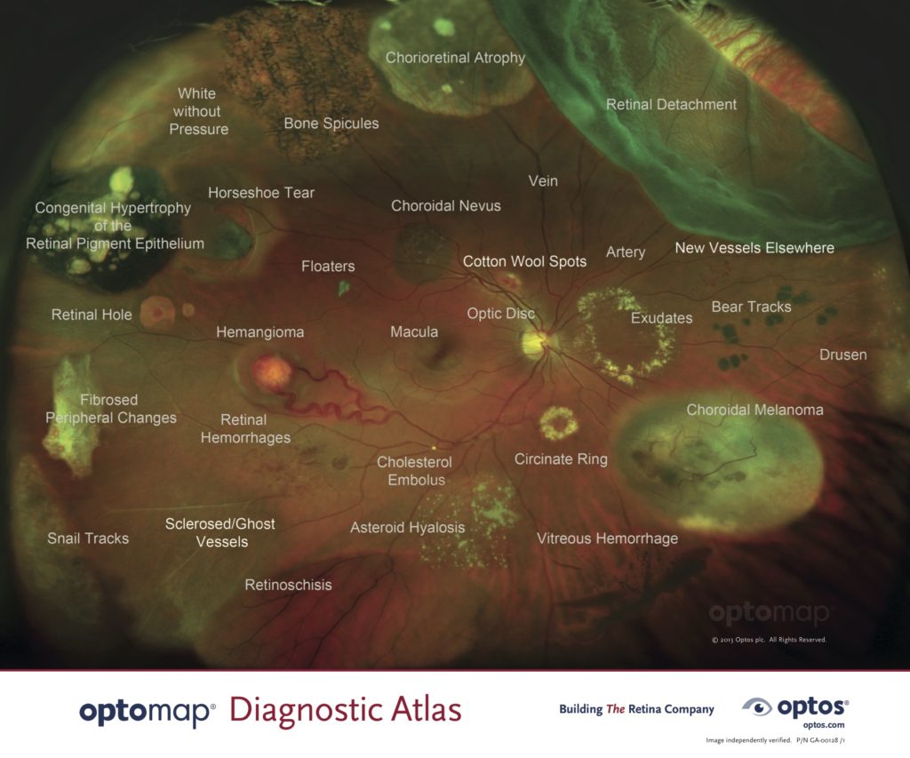
Optomap Policy for Lifetime Eye Care
Our practice takes Optomap images during every vision exam. This is a quick service that provides a better look at your internal eye health, without dilating your eyes. Although this test will add an out-of-pocket cost to each patient, we feel strongly about the benefits of this type of imaging each year. The patient cost is only $29, which is lower than most every other office that offers this service.
Why is it so important?
With Optomap images, we are able to evaluate retinal health to a much greater extent and have an image to compare to in the future. Although most of the time the images show a healthy retina, we have found problems ranging from minor to severe in hundreds of patients, and many times without any warning or symptoms. The retina is the light-sensitive portion at the back of the eye that transmits the signals of light from the world to your brain so you can see. This is the part of the eye that can suffer from macular degeneration, glaucoma, diabetic retinopathy, melanoma, retinal holes/detachments, genetic disorders, and several other conditions. Vision loss can result from these conditions, many of which are treatable and vision loss may be prevented with early detection.

How does it work?

Taking Optomap images is quick and easy, and no eye dilation is required. It just takes a few moments during pretesting to line your eye up in the machine until you see the green light, then a quick 1-2 second bright flash of light, and the image is ready. If you blinked, then we just retake it. We obtain a couple of images of each eye. The machine uses green and red laser to image the different layers of the retina. The doctor will review your images with you in the exam room. If any problems are detected, the doctor will let you know if it requires a referral, treatment, further testing, or monitoring.
FAQ
Why don’t insurance companies pay for this test? This is still considered an advanced procedure that most offices in the country don’t yet have.
Is this just about money? No, it’s about a thorough test of eye health and detecting problems that may exist. We are keeping the cost low because we are sensitive to the extra fee, but as a very expensive piece of equipment, a fee is necessary.
Can I opt-out of this service? No, we are making this our standard of care for all patients during every vision exam.
What about kids? Yes, we also take Optomap images of children. Kids as young as 4 years old are usually able to do the test well. We have found serious problems in healthy young children with no complaints, so testing is important for them as well. If your child is physically unable to take photos there will obviously be no charge.
Will I need to have my eyes dilated? No, in almost every case, Optomap images allow us to see your internal ocular health without dilation during your annual vision exam. Please note that we see many patients for medical eye exams for diabetes, cataracts, macular degeneration, and other conditions. Many of these exams still require dilation, which is typically done at a medical eye exam separate from an annual vision exam.
Does this policy apply to medical eye exams? No, this policy applies to vision exams, which provide an evaluation of each component of vision. Medical eye exams are more problem-focused and may include dilation and retinal photos when necessary, oftentimes which can be billed to medical insurance when an underlying condition is present.
What if there is nothing wrong? Great! In most cases, we are happy to find normal ocular health, but we don’t know if there is a retinal problem until we look. The images are still very useful as we can use them as a comparison in case something changes with your retinal health in the future.
What if I cannot afford the cost? Please let a staff member know if this is your situation.
